Spondylolisthesis Relief in Dallas — Stabilize & Strengthen Your Spine
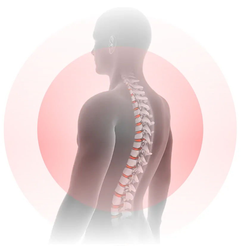
Spondylolisthesis is a forward slip of one vertebra over the next. In younger patients it often stems from stress fractures; in adults it typically develops from disc and facet wear. Left untreated, it can pinch nerves, cause leg pain, and disrupt daily activities.

Low-back pain
worse with
standing
Tight
hamstrings
Sciatica-like pain
down one or both legs
Step-off deformity
seen on imaging
Medications to reduce pain & inflammation
Core strengthening, hamstring stretching, and posture training
Bracing to support alignment and reduce pain
Targeted injections if nerve irritation persists (epidural or facet)
Minimally invasive TLIF/XLIF fusion or motion-preserving implants for severe or progressive cases
Request Your Same-Day Spondylolisthesis Evaluation
Don’t wait in pain — our expert spine specialists are available for same-day evaluations.
A forward slip of one vertebra over another, most often from stress fractures in youth or degenerative changes in adults. See Back & Sciatic Conditions.
Yes—most mild cases improve with medication, therapy, and bracing. See Physical Therapy & Rehab.
Yes—braces can reduce pain and limit progression in early slips. Severe cases may need surgery. See Non-Surgical Therapies.
Fluoroscopy-guided injections can relieve nerve pain and improve mobility when conservative care isn’t enough. See Epidural Steroid Injections.
Fusion is considered for severe slippage, progressive instability, or worsening neurologic symptoms. See Spinal Fusion Surgery.
Some stress-fracture types may run in families, but degenerative cases are usually related to age and wear. See Degenerative Disc Disease.