Arm & Shoulder Injury Specialists in Dallas

Strain, bursitis, arthritis
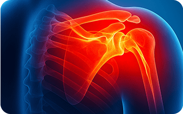
Night pain, weakness; surgical & biologic care

Overuse tendon pain with fast-track relief

Numbness, tingling; bracing, injections, release
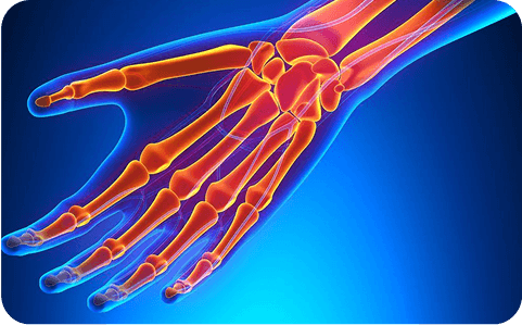
From texting thumb to fractures
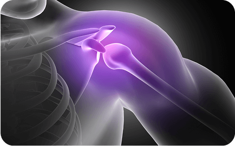
Trauma-related nerve injuries
Explore Other Arm &
Shoulder Diagnoses
Not seeing your condition? Browse our A–Z: arthritis, frozen shoulder, labral tears, nerve entrapments, tendonitis, fractures, and more.


Why Choose Dallas Spine for Arm & Shoulder Care
Board-certified orthopedic spine & pain doctors
Early, accurate diagnosis—no wasted weeks
Personalized, non-opioid-first plans
Fluoroscopy-guided injections for precision
Same-/next-day access at seven DFW clinics
Integrated pathway from therapy to surgery


Request Your Same-Day Arm & Shoulder Evaluation
Don’t wait in pain — our expert spine specialists are available for same-day evaluations.
If you had a fall, crash, or sports injury and lost motion or strength, get imaged now. X-ray rules out fracture; MRI shows tendon or labral tears. Early scans prevent delays in recovery. See Rotator Cuff Tear
No. Many partial/small tears improve with therapy, cortisone, or PRP. Larger/full-thickness tears often need arthroscopic repair. Learn more on Shoulder Pain and Rotator Cuff Tear.
Yes. Wrist trauma can cause swelling that compresses the median nerve—numbness, night pain, grip weakness. Early care allows bracing, injections, or release if needed. See Carpal Tunnel Syndrome.
If pain limits motion, follows trauma, or includes numbness/weakness, get seen. Our board-certified specialists examine, medicate appropriately, and order imaging when needed. Fast diagnosis prevents long-term issues. Start at our Arm & Shoulder Hub.
Earlier evaluation = correct imaging, fewer setbacks, and a plan tailored to you. It reduces scar tissue and speeds return to work and sport. See our Therapies page for non-surgical options.
Yes. Splints stabilize irritated tendons and nerves—useful for carpal tunnel and tendonitis—reducing pain while tissue calms and heals. See Wrist & Hand Pain